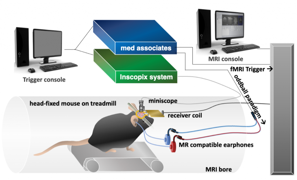
Using functional magnetic resonance imaging (fMRI), studies have provided significant insights into unique large-scale functional organizations of the brain including default mode network (DMN) and salience network (SN). The aberrant communications of brain functional connectivity among these networks have been observed across different classes of addiction and are associated with craving and relapse. The overarching goal of this project is to identify the functional role of anterior cingulate cortex (ACC) in network integration in normal and addiction conditions. Our central hypotheses are that: 1) distinct functional clusters of neurons exist in rodent ACC and these clusters coactivate with distinct large-scale brain networks, 2) SN-related neurons in ACC will be activated in attention tasks whereas DMN-related neurons in ACC will be deactivated. 3) functional integration of SN-related neurons in ACC is decreased in nicotine addiction and causally relates to attention deficits via large-scale brain network dysfunctions. While fMRI can be utilized to depict large-scale brain networks, one technical challenge towards the testing of these hypotheses is to obtain spatially resolved neuronal activity within the ACC during fMRI. To address this, we will utilize an innovative MRI-compatible fluorescent miniature microscope, allowing concurrent imaging of ground-truth neuronal activities at cellular resolution during fMRI. We also implemented a silent zero-echo-time (ZTE) fMRI technique, enabling awake rodent imaging in a stress-free condition. Addressing these hypotheses will clarify the utility of ACC in predicting addiction related attention deficits and its putative role in serving as a novel treatment target for addiction.