MR-compatible multi-channel microelectrode array
Functional magnetic resonance imaging (fMRI) plays a pivotal role in brain research due to its ability to non-invasively acquire whole-brain dynamics with a relatively high throughput. Despite tremendous research efforts, fMRI offers mainly hemodynamic information, which by itself is insufficient to gain mechanistic insights into brain circuit functions. To fill this gap, our group has developed a MR-compatible multi-channel microelectrode array that allows simultaneous fMRI and electrophysiological recordings or micro-stimulation.
a
b
c
d
Our multi-functional microelectrode offers several unique features, including:
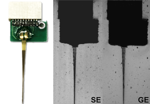
- negligible MR susceptibility artifacts
- robust stimulation and recording capabilities during fMRI
- mechanical flexibility to accommodate MR coils and small gradient inserts
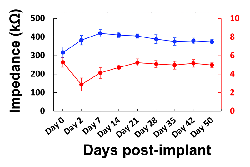
- biocompatibility for longitudinal studies
fMRI with deep brain stimulation
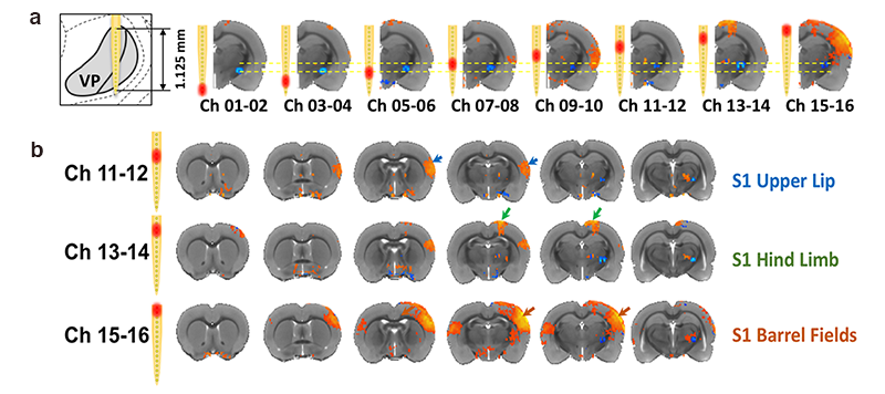
fMRI with simultaneous deep brain stimulation (a) A signature study showing measurable local fMRI responses (blue) from site-selective micro-stimulations of two adjacent channel pairs within the ventroposterior (VP) thalamic complex. (b) Distinct cortical activation patterns from site-selective micro-stimulation within the VP thalamus indicate that our microelectrodes, combined with fMRI, can modulate and map circuit projections at unprecedented sub-nucleus resolution (in 75 μm increments).
fMRI with electrophysiology
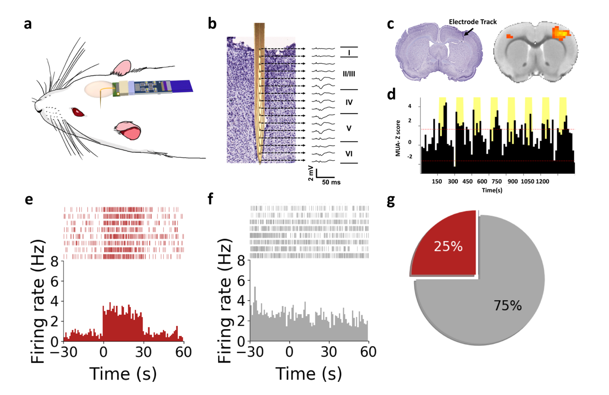
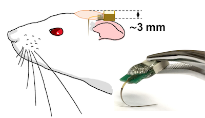
fMRI with concurrent electrophysiology. (a) Schematic setup cartoon. (b) A Nissl stained slice showing microelectrode placement in the somatosensory cortex, alongside the laminar-specific local field potential profiles induced by sensory stimulation. (c) Negligible susceptibility artifact preserves fMRI activation. (d) Multi-unit activity data showing robust responses to each stimulation train. (e) Raster plots and peri-stimulus-histograms of representative neurons showing excitation or no change to stimuli, selected from 16 identified single units in this experiment.
Specification
Microelectrode
Type
Type
Total
Implantable Shank
Implantable Shank
Target
Impedance
Impedance
16-channel linear
15mm
0.5-1.5 MOhm
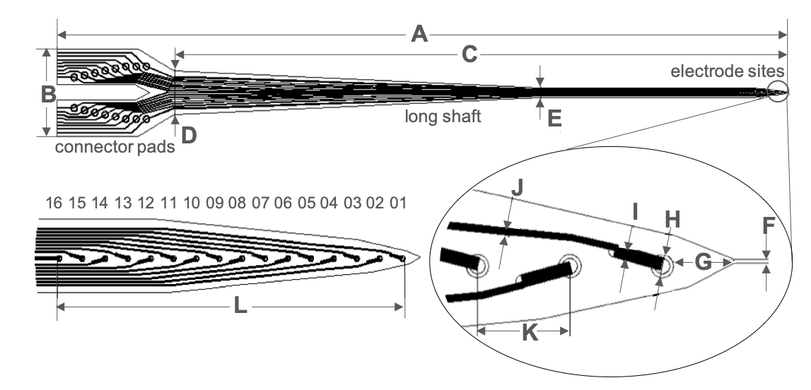
Probe length (A)
18.8mm
Probe width (B)
2mm
Probe length (C)
14.9mm
Max shaft width (D)
996 μm
Shaft width (E)
220 μm
Tip width (F)
3 μm
Distance (G)
48.4 μm
Diameter of electrode (H)
12 μm
Wire width (I)
10 μm
Wire width (J)
5 μm
Electrode distance (K)
Type Ⅰ- 74 μm
Type Ⅱ- 150 μm
Type Ⅱ- 150 μm
Recording length (L)
Type Ⅰ- 1.11mm
Type Ⅱ- 2.25 mm
Type Ⅱ- 2.25 mm
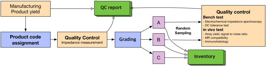
Product quality management and User Training
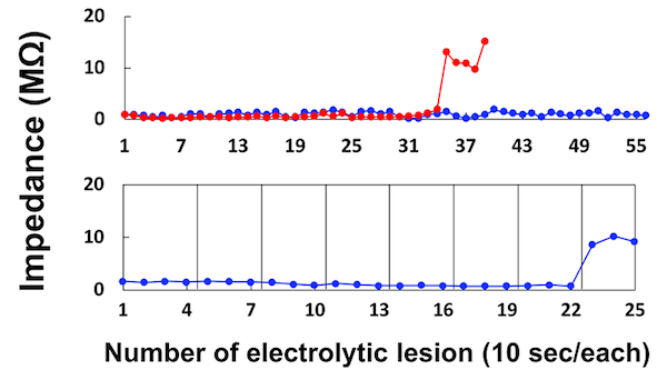
Quality Control: Our goal is to operate commercial grade quality control for our microelectrodes as shown below.
User Training: We are preparing user training materials including a tutorial video and step-by-step instructions. In the meantime, please contact camri@unc.edu if you have any questions about using our microelectrodes.
How to Order
Due to the technical nature of microelectrode production and quality control, and limited budget, we can only accommodate limited requests for our microelectrodes (about 2 electrodes per research group). We hope to scale up our production in the future.
If you are interested in our products or have any questions, please contact camri@unc.edu.