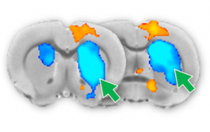
Our understanding of human brain function has been heavily influenced by functional magnetic resonance imaging (fMRI) – a technique that measures neuronal activity indirectly through blood oxygenation changes. Accumulating data indicates that our interpretation of fMRI data could have been wrong, or even opposed to the actual neuronal processes in a brain area called striatum. The striatum is involved in cognition, motivation, reward, sensorimotor function, and several neurological and neuropsychiatric disorders such as addiction, obsessive-compulsive disorder, schizophrenia, Parkinson’s disease, and major depression. In this project, we plan to use cutting-edge neuroscience tools to stimulate and record cellular activity as well as neurotransmitter release in the striatum during fMRI to uncover the mechanism by which fMRI signal is formed in the striatum.
